Release date: 2007-08-14 The 256-row new CT scanner has a 256-row CT scanner equipped with the world's most advanced CT imaging software and devices at the Johns Hopkins School of Medicine in North America. It has been a three-month safety and Clinical evaluation.
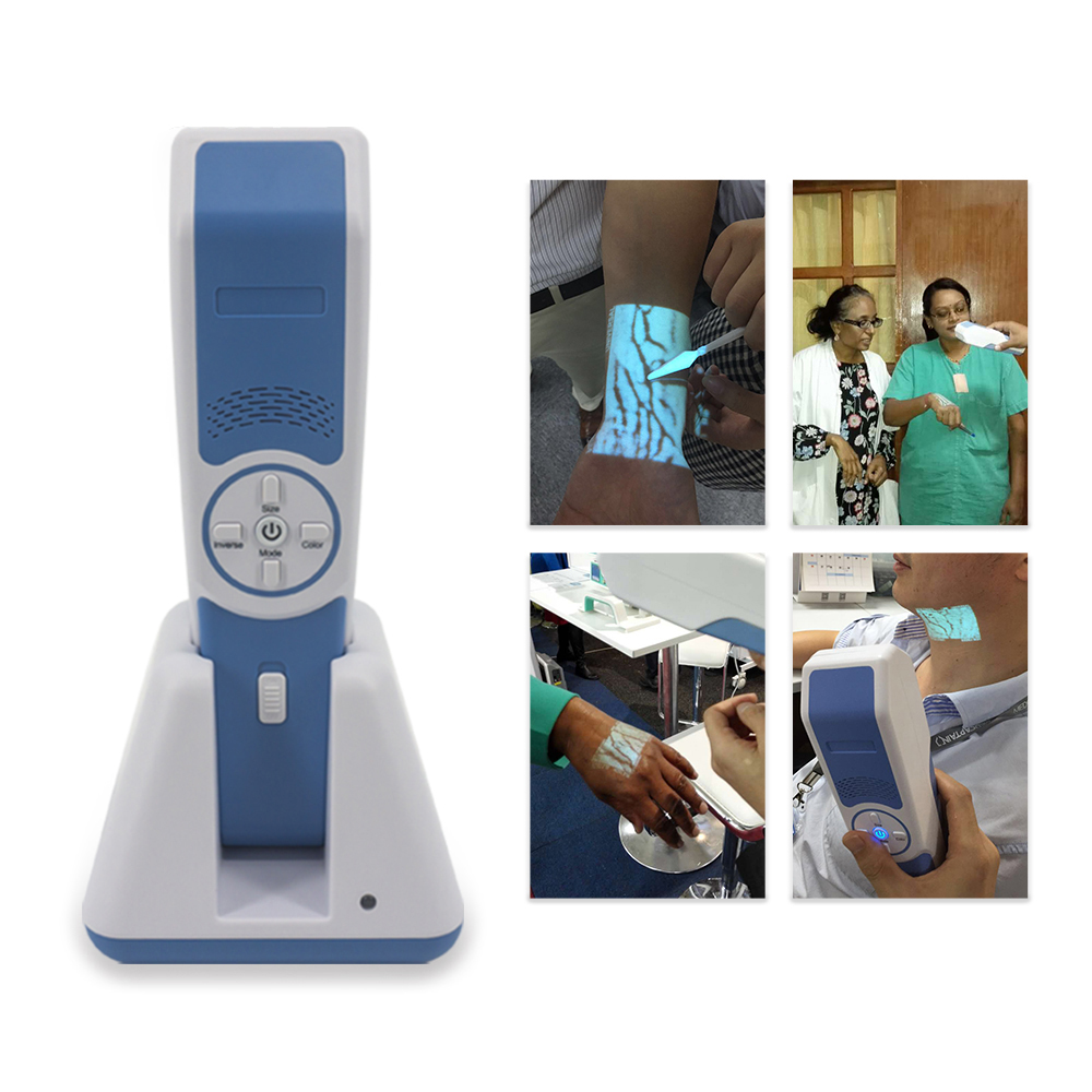
vein finder bases on the near-infrared absorbance difference between veins and surround tissue, it projects accurate and real-time vein image on the skin of patient. vein finder can be used to find and evaluate the patients` veins, help nurse to do vein puncture. vein finder can help increasing first vein puncture attempt successful rate, increasing vein finding and puncture efficiency, increasing patients` satisfaction and decrease medical disputes. vein finder is widely used in the following departments: pediatric, emergency, oncology, geriatric, radiology, laboratory, vascular surgery, plastic surgery, etc.
Infrared Vein Finder Infrared Vein Finder,Handheld Infrared Vein Finder,Medical Infrared Vein Finder Vein,Medical Infrared Vein Finder Shenzhen Guangyang Zhongkang Technology Co., Ltd , https://www.lighttherapymachine.com
This new device is the first in North America. In addition, there is only one in Japan, which is four times the coverage of its predecessor, 64-slice CT. It can detect fine changes in blood flow or no more than 1.5 mm in the heart and brain. A tiny thrombus formed in the blood vessels of a toothpick. Production producers say that Aquilion beta 256, produced by Toshiba Corporation of Japan, is expected to be approved for a wide range of clinical applications within one year. Hopkins Medical School is negotiating the price of the device, which is priced at $1 million.
Dr. Jo?o Lima, a cardiovascular specialist at the Johns Hopkins School of Medicine who is responsible for all cardiovascular testing, said the device has the advantage of being able to detect the most restrictive blood flow before symptoms occur or before permanent organ damage occurs. Signs. Lima, an associate professor at the Johns Hopkins School of Medicine and the Heart Institute, said that arterial, venous, or capillary blockage in any organ can develop slowly for many years, with chest pain, severe fatigue, and headache symptoms only when the disease is severe enough to threaten life. The key technological advances of the 256-CT (it seems that the patient table is surrounded by a giant ring-shaped metal ring, called the central frame) is the number of probes, each probe scanning range is four times that of 64-CT. Hopkins Medical currently has a 64-row CT scanner.
According to the company, the X-ray emission structure of the device can form a 12.8 cm (5 inch) diameter single shot, and the thickness of the single layer is sufficient to cover most single organs at one time, including the brain, heart, intact joints, and most of the lungs. And the liver. This is an improvement in 64-slice CT 3.2 cm imaging coverage, and 64-slice CT requires several rotations or scans to complete the visualization of a single organ. Dr. Kieran Murphy, an international neuroradiologist and associate professor of radiology at Hopkins University, said he believes that the entire head perfusion imaging scan can detect areas of the brain that are prone to stroke and have a slow blood flow, with a single scan.
CT imaging consists of X-rays that penetrate the body to produce detectable digital signals and computer reconstruction. Each of the 256 probes in the new device captures a layer of organ or tissue signals. The more probes, the better the image will be. The computer combines all layer signals and synthesizes a detailed three-dimensional image of the heart or brain and surrounding blood vessels. In some cases, the patient is injected with developer to add more detail. Murphy, who is responsible for the neurological detection of the scanner, said that the extended coverage has a huge advantage over older machines. Older machines require matching and overlapping imaging, and over time, similar to the complex technical procedures for rebuilding multiple layers of marble on each layer. He also mentioned that the cooling system no longer needs to deal with the friction and heat caused by multiple rotations of the structure, although computer software still requires a cooling system. Murphy said that the data output per scan of the 256-CT increased to 5-10 billion bytes, while the 64-CT has only 1 to 2.5 billion bytes.
However, he does not expect higher data capacity to slow down the check, 256-CT acceleration for at least 1 second for brain imaging, and 64-CT for 4 or 5 seconds. Lima said that the total time required for a 256-CT cardiac exam is also reduced to 1 or 2 seconds, while 64-CT requires 8 or 10 seconds. Lima said that the new and fast device will also make it possible to scan patients with arrhythmia or irregular heart rhythm. The 256-CT completes all imaging in only one heartbeat interval, while the 64-CT requires longer time----6 or 8 heartbeat intervals. He said that any interference between continuous heartbeats, such as those produced by arrhythmia, can cause the synthesized scanned image to distort. A single scan of 256-CT can also provide all five important cardiac diagnostic tests or single brain tests for patients with the most severe conditions, making them less exposed to radiation, about 1/8 of the dose required for a 64-row CT scan. To 1/3. Since a single scan of 256-CT should provide patient calcification, blood flow data, heart rate, and pulse intensity for examining arterial stiffness, it is more accurate for more invasive procedures (such as cardiac catheterization or brain angiography). Optimal patients expect value.
The new scanner and nine technicians were loaned to Hopskin from Toshiba America Medical Systems. The temporary installation and renewal costs of Hopskin are also borne by Toshiba. —— Information from: Shanghai Medical Device Industry Association 
256-row new CT scanner operating in North America
Prev Article
Canned Asparagus